X-rays use invisible electromagnetic energy beams to produce images of internal tissues, bones, and organs on film or digital media. Standard X-rays are performed for many reasons, including diagnosing tumors or bone injuries .
FUJIFILM offers X-ray equipment, films, printers, and solutions.
| X-RAY EQUIPMENT
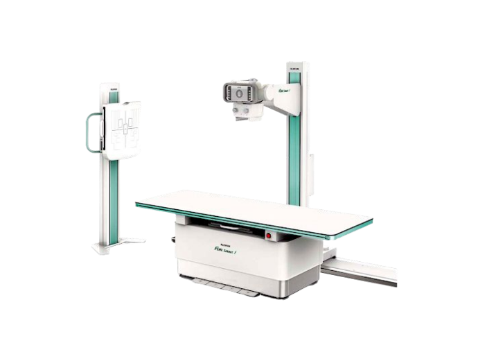
FDR SMART X
Fujifilm’s Advanced X-Ray System
Learn More
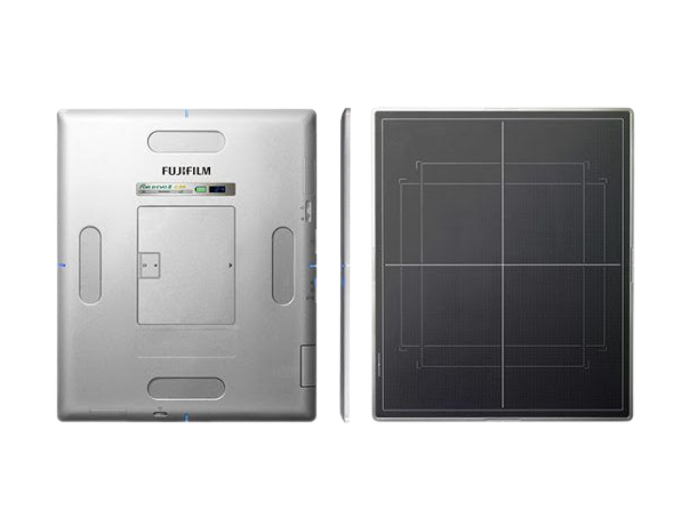
FDR D-Evo II
Fujifilm’s Flat Panel Detector
Learn More
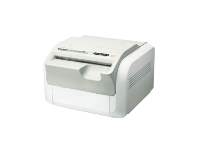
FCR Prima T2
Table-top Digital Image Reader
Learn More
| X-RAY PRINTERS
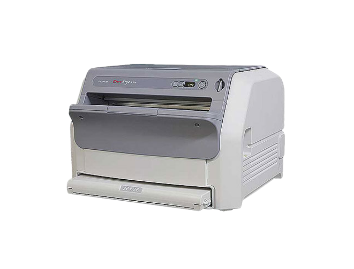
DRYPIX Lite
Medical Dry Thermal Film Printer
Learn More
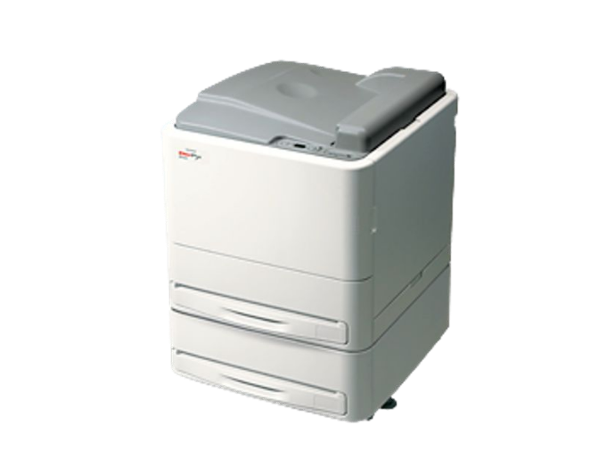
DRYPIX Smart
Medical Dry Laser Film Printer
Learn More
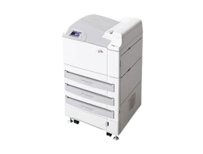
DRYPIX Plus
Medical Dry Laser Film Printer
Learn More
| X-RAY FILMS & CASSETTES

Super HR-U
Conventional X-Ray Films
Learn More

DI-HL
Dry Imaging Laser X-Ray Films
Learn More

DI-HT
Dry Imaging Thermal X-Ray Films
Learn More

FCR Imaging Plates and Cassettes
For High Quality Digital Imagess
Learn More
| X-RAY SOLUTIONS

FUJIHUNT XF-25 X-RAY Fixer
For manual processing of all X-Ray Films
Learn More

FUJIHUNT Automatic X-RAY Fixer
For the automatic processing of all X-Ray Films
Learn More

FUJIHUNT XD-25 X-RAY Developer
For manual processing of all X-Ray Films
Learn More

FUJIHUNT Automatic X-Ray Developer
For the automatic processing of all X-Ray Films
Learn More
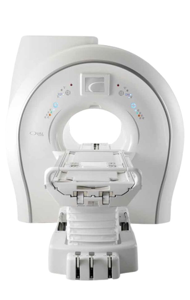
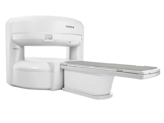
Magnetic resonance imaging (MRI) is a medical imaging technique that uses a magnetic field and computer-generated radio waves to create detailed images of the organs and tissues in your body. MRI scanners are particularly well suited to image the non-bony parts or soft tissues of the body.
Computed tomography, also known as CT scan, is used to examine structures inside your body. A CT scan uses X-rays and computers to produce images of a cross-section of your body. It takes pictures that show very thin “slices” of your bones, muscles, organs and blood vessels so that healthcare providers can see your body in great detail.
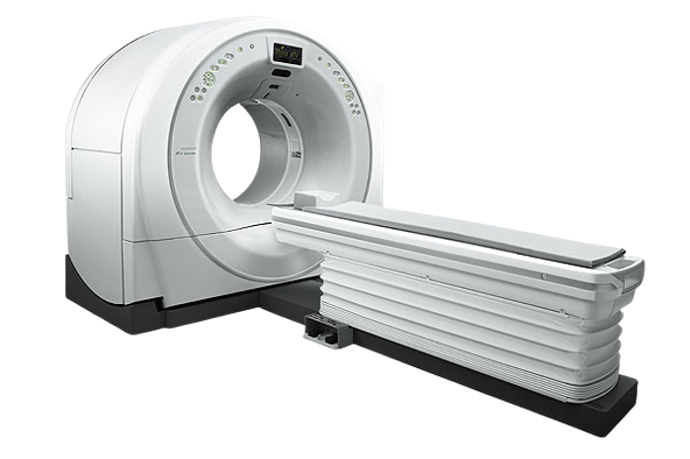
FCT Speedia HD
Compact CT Device with a High Speed Scan
Download Brochure
Diagnostic ultrasound, also called sonography or diagnostic medical sonography, is an imaging method that uses high-frequency sound waves to produce images of structures within your body. The images can provide valuable information for diagnosing and treating a variety of diseases and conditions.
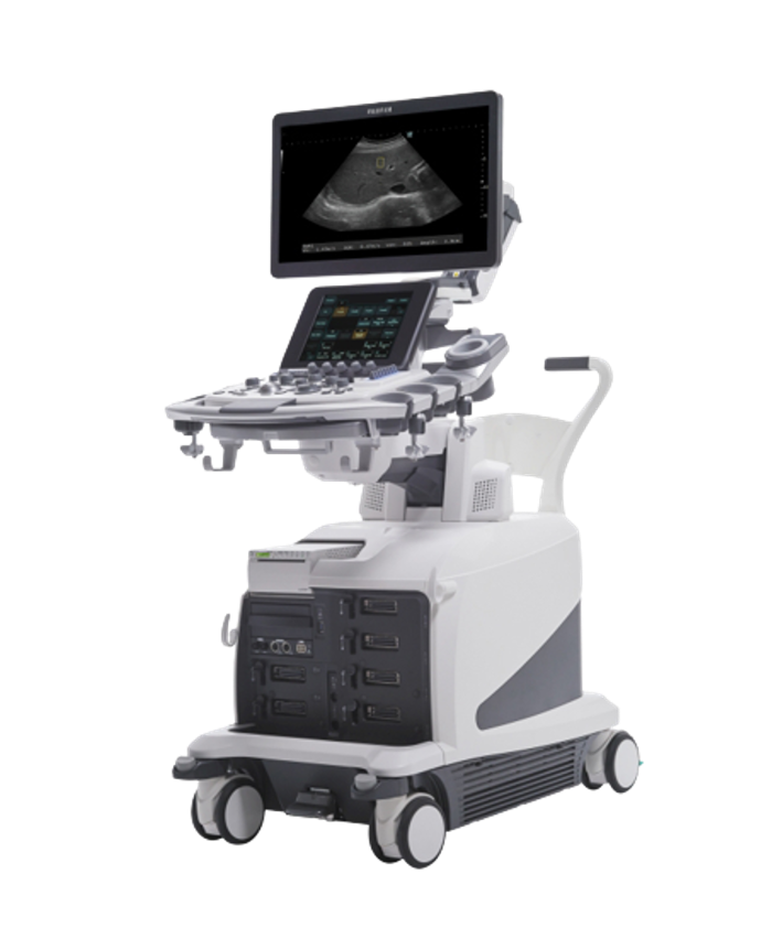
ARIETTA 850
Ultrasound Scanner for Radiology
Download Brochure
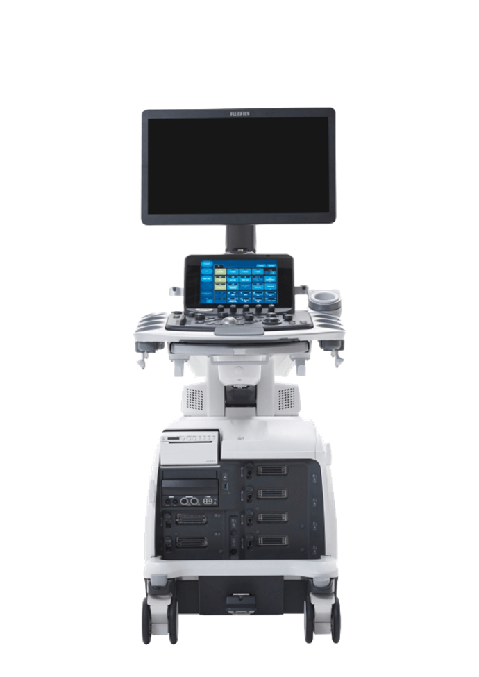
LISENDO 880
Ultrasound for Cardiology
Download Brochure
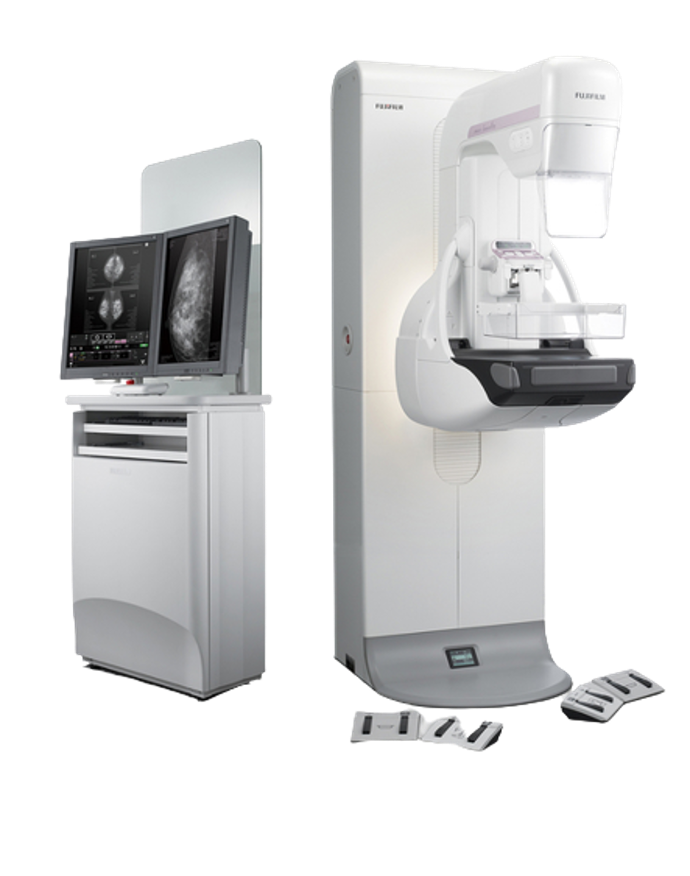
Mammography is the process of using low-energy X-rays to examine the human breast for diagnosis and screening. The goal of mammography is the early detection of breast cancer, typically through detection of characteristic masses or microcalcifications.
